
Digital radiology in Sant Just Desvern
More accurate diagnoses for safer treatments
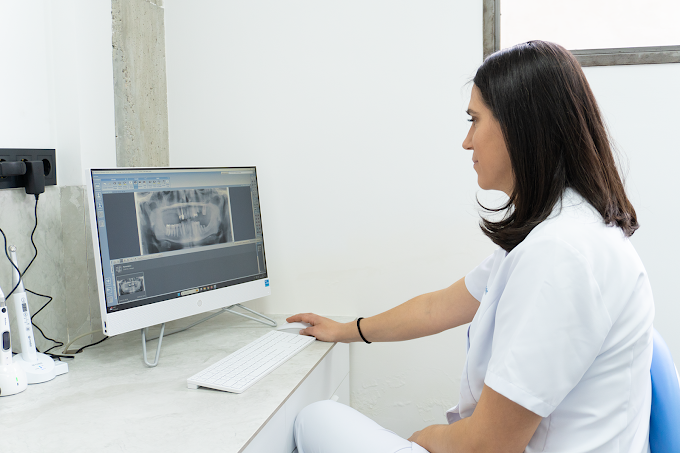
Accuracy and reliability
Modern dentistry has evolved tremendously thanks to technological advances in diagnostic imaging. Today, many pathologies and clinical situations that previously went unnoticed can be detected with great precision thanks to digital radiology systems. At VARCHI Dental Clinic, we understand that quality treatment always begins with an accurate diagnosis. Therefore, we utilize 2D and 3D digital radiology technology, allowing us to obtain detailed images of teeth, gums, jawbones, and other anatomical structures of the oral cavity.
These tools help us plan treatments more safely, predictably, and in a more personalized way, reducing uncertainties and improving clinical accuracy across all dental specialties. Radiology is not a treatment in itself, but a fundamental tool for understanding what lies beyond the visible surface. Many dental diseases, infections, bone loss, or anatomical abnormalities can only be accurately detected through appropriate imaging tests.
Thanks to current digital technology, we can obtain extremely precise information using controlled radiation doses tailored to each patient's needs. Our goal is to provide reliable diagnoses that allow us to make clinical decisions based on detailed and objective information.
What is digital dental radiology?
Digital dental radiology is the set of diagnostic imaging techniques used to visualize internal structures of the oral cavity that cannot be observed during a conventional clinical examination. These images allow for the highly precise analysis of teeth, roots, jawbone, gums, and other anatomical structures.
The main advantage of digital radiology over traditional systems is the image quality, the speed of obtaining results, and the possibility of performing much more detailed analyses.
Furthermore, digital technology allows for optimized radiation doses and significantly improved diagnostic capabilities.
The information obtained is essential for planning treatments in general dentistry, implantology, oral surgery, orthodontics, endodontics, and periodontics, among other specialties.
Solutions
Accurate diagnosis from the start
In modern dentistry, clinical diagnosis and radiological diagnosis work together to provide a complete view of oral health.
2D Radiology
Two-dimensional radiographs remain a fundamental tool in daily dental diagnosis. They allow for quick and precise imaging of different structures in the oral cavity and are a routine part of clinical examinations and evaluations. Depending on the information we need, we use different types of radiographs.
Orthopantomography (Panoramic X-ray)
An orthopantomogram, also known as a panoramic radiograph, allows visualization of all teeth, both jaws, the temporomandibular joints, and other relevant anatomical structures in a single image. It is one of the most widely used tests in dentistry due to its ability to provide a comprehensive view of the mouth. It is especially useful for assessing tooth position, detecting missing teeth, identifying impacted wisdom teeth, evaluating bone loss, and conducting initial studies for numerous treatments. It is also a fundamental tool in oral surgery, implantology, and orthodontics.
Periapical radiographs
Periapical radiographs provide highly detailed images of one or more specific teeth, including their roots and the surrounding bone. Their high definition makes them an indispensable tool for diagnosing deep cavities, infections, root fractures, and problems related to endodontic treatments. They also allow for monitoring the progression of certain pathologies and evaluating the results of previously performed treatments.
Interproximal radiographs or bitewing radiographs
Bitewing radiographs are particularly useful for detecting cavities between teeth that are not visible during a conventional examination. They also allow for the evaluation of the supporting bone and the monitoring of certain periodontal conditions. They are a very useful tool within prevention and early diagnosis programs.
3D digital radiology: CBCT technology
In certain clinical situations, we need to obtain much more detailed information about anatomical structures. For this, we use CBCT (Cone Beam Computed Tomography) technology, an advanced form of computed tomography designed specifically for dentistry. Unlike conventional X-rays, CBCT generates three-dimensional images that allow us to visualize teeth, bones, and anatomical structures from any angle. This three-dimensional view provides an extraordinary level of diagnostic accuracy and is especially useful in complex treatments.
Instructions
Why is it important to perform radiological tests?
Many dental pathologies progress without symptoms. Radiological tests allow them to be detected before they become clinically visible.
Radiological safety and control
Patient safety is an absolute priority in any diagnostic procedure. Therefore, we only perform those tests that are truly necessary for diagnosis and treatment planning.
We work following strict radiation protection protocols and use modern technology designed to optimize image quality with the least possible exposure.
Each examination is indicated on an individual basis, carefully assessing the diagnostic benefit obtained. Our commitment is to combine diagnostic accuracy and safety in every procedure performed.
Protocol
Applications of digital radiology in dentistry
Radiology is part of virtually all dental specialties. Each image obtained contributes to a more accurate diagnosis and a more personalized treatment plan.
General dentistry
In general dentistry it helps to detect cavities, infections and dental abnormalities.
Periodontology
In periodontics, it allows for the evaluation of the condition of the bone that supports the teeth and the assessment of the evolution of periodontal diseases.
Implantology
In implantology, it provides essential information for planning the safe placement of implants.
Oral surgery
In oral surgery, it facilitates the planning of complex procedures and reduces surgical risks.
Orthodontics
In orthodontics, it allows the study of the patient's dental and skeletal development.
Root canal treatment
In endodontics, it provides detailed information about roots, canals, and periapical tissues.
Advantages
Advantages of digital radiology
The incorporation of digital technology has brought about a true revolution in dental diagnosis.
01/
Higher image quality
It allows for sharper and more detailed images than conventional systems.
02/
Analysis and monitoring
Images can be enlarged, reviewed, and stored digitally to facilitate clinical monitoring.
03/
Immediate results
Obtaining images is quick, speeding up diagnosis and explanation to the patient.
04/
Greater security
It allows for optimizing the radiation dose while maintaining high diagnostic quality.
FAQs
Your dental practice
Why choose VARCHI Dental Clinic for your radiological diagnosis?
At VARCHI we are committed to dentistry based on precision, planning and technology applied to patient care.
Book your appointment at VARCHI
We’ll assess your situation and clearly explain all the options available to help you regain your smile.